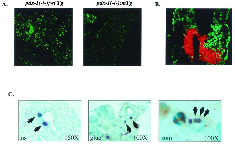
Figure 4.
Pancreata from pdx-1 (−/−);mTgt mice show a defect in proliferation and contain increased numbers of hormone-positive cells in the ductal epithelium. (A) Immunocytochemical staining of whole-pancreas sections from pdx-1 (−/−);wtTg and pdx-1 (−/−);mTg neonates, with Ki-67 antiserum as the marker of cell proliferation. (B) Immunocytochemistry of pancreas sections from neonatal wild-type mice, showing the highest levels of PBX-1 (green) in ductal cells (D), followed by islet cells. Acinar cells stain weakly for PBX-1. Insulin immunostaining (red) is overlaid to indicate the islet. (C) Immunohistochemical detection of insulin (Left), glucagon (Middle), and somatostatin (Right) cells (arrowheads) within the pancreatic ductal epithelium of neonatal pdx-1 (−/−);mTg mice. Mice of all other genotypes did not show hormone-positive cells within ducts in any sections examined.

