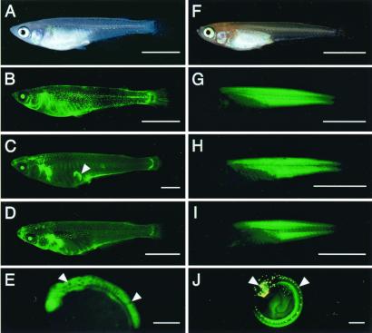
Figure 1.
Expression of the GFP in nuclear transplants and their offspring. (A) A 1.5-month-old donor transgenic fish, carrying EF-1a-A/GFP, under visible light. The body color is that of the wild type. (B) A fluorescent image of A. An intense fluorescence in the eyes and a weak one throughout the skin is observed. Fluorescence in the belly and a dot-like one in the trunk are autofluorescence of pigment cells. (C) A 7-month-old nuclear transplant (1NT1). The arrowhead indicates an artifact. (D) A 1-month-old F1 offspring derived from 1NT1. (E) A 1.5-day-old embryo from an F2 offspring derived from 1NT1, with the wild-type body color showing densely pigmented melanophores in the embryonic body (arrowheads). (F) A 3-week-old donor transgenic fish, carrying β-Act/GFP-N, under visible light. The body color is orange-red. (G) A fluorescent image of F. An intense fluorescence is observed in the muscle tissue. (H) A 3-week-old nuclear transplant produced in a separate experiment. In the second experiment, no photograph of the nuclear transplants was taken to avoid accidental killing of the fish. (I) A 1-month-old F1 offspring derived from 2NT1. (J) A 5-day-old embryo from an F2 offspring derived from 2NT4. Yellow spots in the head and on the dorsal midline are autofluorescence of leucophores (arrowheads). [Bars = 5 mm (A–D and F–I) and 0.3 mm (E and J).]

