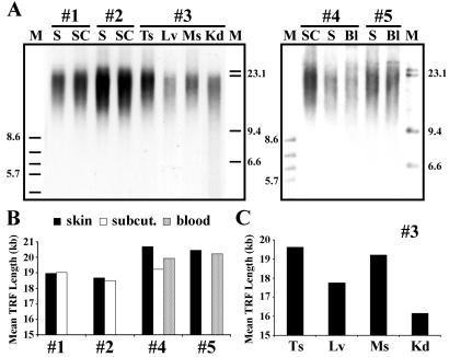
Figure 5.
Variation of telomere lengths between cloned calves and among different tissues within each cloned animal. (A) TRF analysis of digested DNA from various tissue samples of multiple clone calves (#1–#5; <3 weeks old) derived from the same bovine FF cell line. Samples are skin (S), s.c. (SC), testis (Ts), muscle (Ms), kidney (Kd), and blood (Bl). Molecular weight markers (M) are in kilobases. (B) Densitometric analysis of TRF profiles revealing mean TRF lengths of different samples (skin, s.c., and blood) from multiple cloned offspring (#1, #2, #4, #5). (C) Mean TRF lengths in testis (Ts) (mean TRF length = 19.63 kb), liver (Lv) (mean TRF length = 17.76 kb), muscle (Ms) (mean TRF length = 19.23 kb), and kidney (Kd) (mean TRF length = 16.16) tissues from clone #3.

