Figure 2.
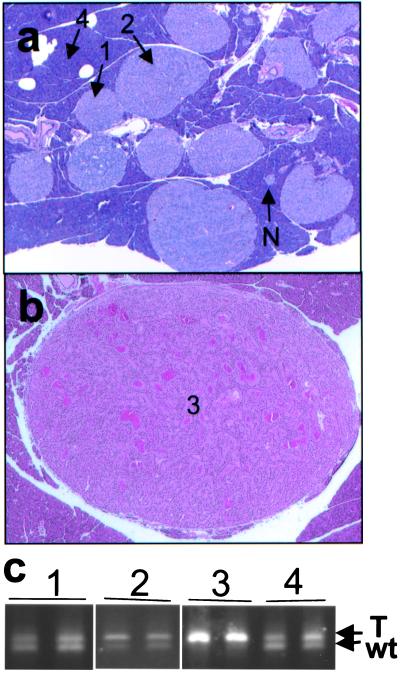
Pancreatic lesions in Men1TSM/+ mice. (a) H&E-stained section (×15) of pancreas from a 12-month-old mouse, showing normal islets (N), hyperplastic islets (1), hyperplastic islets with dysplasia (2), and acinar pancreas (4). (b) H&E-stained section (×15) of a pancreatic islet cell tumor (3) from a 15-month-old mouse, showing increased vascularization and ribbon-like nuclei formation around capillaries. (c) Agarose gel electrophoresis of PCR from microdissected samples. Numbers correlate with the type of tissue indicated in a and b: 1 is hyperplastic islet, 2 is hyperplastic islet with dysplasia, 3 is pancreatic islet cell tumor, and 4 is acinar tissue. The upper band arises from the TSM allele (T), and the lower band from the wild-type allele (wt).

