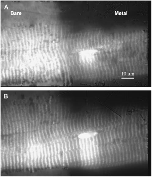
FIGURE 6.
A single skeletal muscle fiber labeled with 5′IATR and observed with fluorescence under metal film-enhanced TIRM (right) and TIRM at a bare-glass interface (left). Metal film thickness is 30–40 nm. Panels A and B correspond to different evanescent field depths. Panel B has the normal depth of ∼100 nm. Panel A is shallower but deeper than the minimal depth in Fig. 8. Observations were recorded on a CCD camera using a rhodamine filter set. The striated pattern is due to 5′IATR localization in the thick filament at the myosin cross-bridge. The bare-glass/metal-film boundary is demarcated by the abrupt change in background fluorescence. The bright oval structure is a nucleus that has scattered surface plasmons creating propagating light that excites fluorescence deeper in the fiber.

