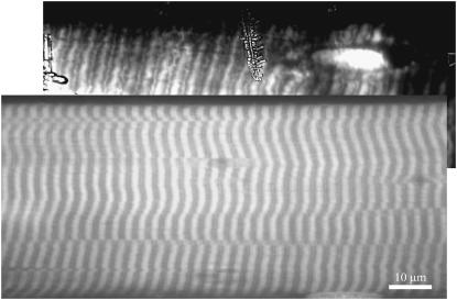
FIGURE 7.
Comparison of fluorescence images of two single skeletal muscle fibers labeled with 5′IATR. The upper image was taken under metal film-enhanced TIRM. Metal film thickness is 30–40 nm. The lower image was taken with a conventional diffraction-limited confocal microscope (no metal film present). Different fibers were used in the two images; however, both fibers were from the same 5′IATR-labeled fiber bundle. The metal film-enhanced TIRM image shows 5′IATR label localized in the Z-disk of the fiber and reveals features of Z-disk structure. The fluorescence probably originates from the small fraction of α-actinin modified by 5′IATR. The TIRM image shows ice crystal formation on the camera window.

