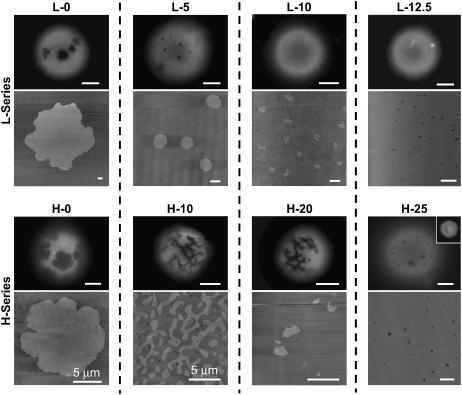
FIGURE 2.
GalCer domain microstructure for slow-cooled supported lipid bilayers and GUVs, an unsupported equilibrium model membrane system. The top set of images corresponds to the L-series compositions (refer to Table 1). Domain size and shape were consistent at all compositions. The disappearance of observable domains at L-10 in the GUVs results from the lower resolution of optical fluorescence microscopy. The bottom set of images corresponds to the H-series compositions. Domain size and shape were consistent at all compositions except H-20 and H-25. At H-20 the domains in GUVs maintained the networked domain microstructures whereas the domains in slow-cooled supported bilayers adopted small irregular shaped ovals. At H-25 GalCer and DLPC became miscible in slow-cooled bilayers whereas in GUVs we observed circular domains that were at the resolution limit of optical fluorescence microscopy. At H-30 no domains were observed in GUVs, indicating lipid miscibility (inset). Fluorescence scale bar 10 μm. AFM scale bar 1 μm, unless indicated otherwise.

