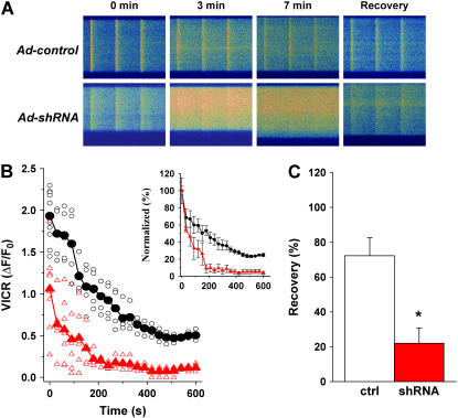
FIGURE 8.
Abnormal maintenance of intracellular Ca2+ storage in skeletal muscle with suppression of JP1 and JP2. (A) Confocal line scan measurement of voltage-induced Ca2+ transients in viral-infected FDB muscle cells (loaded with 10 μM Fluo-4-AM). Time elapsed from the removal of extracellular Ca2+ is indicated. Changes in the transverse dimension of the confocal image are due to variations in fiber shape at different line scan positions. (B) Decline of VICR with time in 0 [Ca2+]o is averaged from multiple experiments (n = 5 for Ad-control, n = 7 for Ad-shRNA). Circles represent individual Ad-control fibers (black); triangles represent Ad-shRNA fibers (red). Normalized VICR with VICR at time 0 is replotted to demonstrate the depletion rate of SR Ca2+ stores (inset panel). (C) After field-stimulated depletion of SR Ca2+ content, 2 mM Ca2+ was restored to the extracellular solution. The recovery of VICR was measured 20 min later (n = 8 for Ad-control; n = 7 for Ad-shRNA). Increasing the recovery time to 35 min did not significantly influence the response in Ad-shRNA muscle fibers. Data are presented as the mean with error bars representing one standard deviation, p < 0.001.

