
Fig. 4.
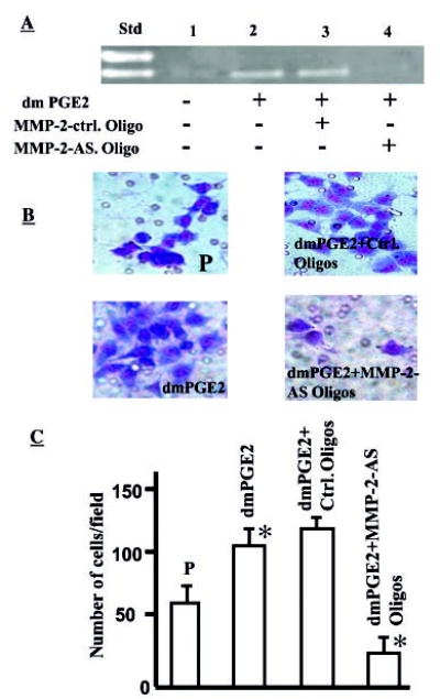
A, PGE2 augments MMP-2 activity in NSCLC cells. SFM was collected from A549-P (P; 106 cells/ml) in the absence or presence of dmPGE2 (10 μg/ml) and run on gelatin zymogram gels. Std, standard; lane 1, A549-P cells in medium alone without PGE2; lane 2, A549-P cells with dmPGE2 (10 μg/ml); lane 3, A549-P cells with dmPGE2 (10 μg/ml) plus MMP-2 control oligonucleotides; lane 4, A549-P cells with dmPGE2 (10 μg/ml) plus MMP-2 antisense oligonucleotides. An increase in MMP-2 level was observed in PGE2-treated cells (lane 2). This PGE2-mediated increase in MMP-2 was effectively blocked by MMP-2-AS oligonucleotides (lane 4). B, PGE2-induced NSCLC invasion is MMP-2-dependent. A549-P cells cultured in medium alone (upper left panel), in the presence of 10 μg/ml dmPGE2 (lower left panel), dmPGE2 plus MMP-2 control oligonucleotide (upper right panel), and dmPGE2 plus MMP-2 antisense oligonucleotide (lower right panel) were used for Matrigel matrix assays. Compared with control (upper left panel), significantly more cells invaded in the presence of dmPGE2 (lower left panel) and fewer cells invaded in the presence of MMP-2 antisense oligonucleotide (lower right panel). C, bar graph representing the number of cells invaded in each group. A 2.5-fold increase in the number of cells invading the matrix is seen in dmPGE2-treated cells (p > 0.05), whereas significantly fewer cells invaded in MMP-2 antisense oligonucleotide-treated cells (p < 0.05).