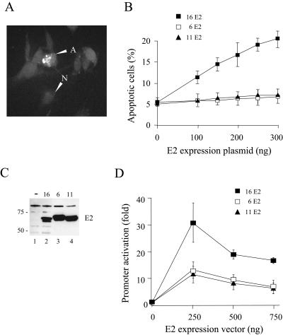
FIG. 1.
LR-HPV E2 proteins fail to induce apoptosis. (A) A plasmid expressing HR-HPV16 E2 was transiently cotransfected into HeLa cells along with a plasmid that expresses RFP (pHcRED-C1). After 30 h, apoptotic cells in the transfected population were identified using a TUNEL assay with fluorescein-12-dUTP. A, apoptotic nucleus; N, normal nucleus. (B) pHcRED-C1 and plasmids expressing HR-HPV16, LR-HPV6, or LR-HPV11 E2 were transiently cotransfected into HeLa cells, and transfected apoptotic cells were counted as described above. The transfection was performed in duplicate and repeated three times, and the data are the means and standard deviations. (C) Cos-7 cells were transiently transfected with plasmids expressing HR-HPV16 E2 (lane 2), LR-HPV6 E2 (lane 3), or LR-HPV11 E2 (lane 4) fused to eGFP or left untransfected (lane 1). After 24 h, whole-cell lysates were separated by SDS-PAGE and transferred to a PVDF membrane. The tagged proteins were detected using anti-eGFP monoclonal antibodies (Covance). The sizes of the markers used are indicated. (D) HeLa cells were transiently cotransfected with an E2 responsive luciferase reporter plasmid and the E2 expression vectors described above. Luciferase activity was normalized for transfection efficiency using a cotransfected plasmid expressing Renilla luciferase. The data are presented as promoter activity relative to the reporter alone and are the means and standard deviations of results from three independent experiments.

