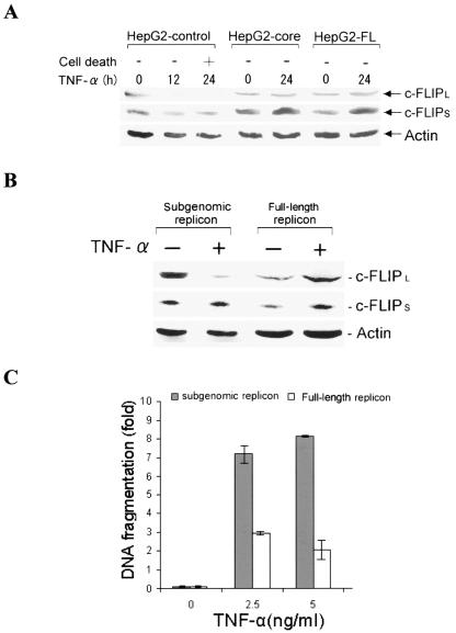
FIG. 4.
c-FLIPL expression level is sustained in TNF-α-treated HCV core-expressing hepatocytes. (A) Western blot analysis for expression of c-FLIP from TNF-α-treated HepG2 cells stably transfected with empty vector (control), HCV core, or FL cDNA. The expression level of c-FLIPL in TNF-α-treated HepG2 control cells could not be detected after 24 h, while its expression was sustained in HepG2-core or HepG2-FL cells. The level of c-FLIPS increased upon TNF-α treatment of HepG2-control, HepG2-core, or HepG2-FL cells. (B) Western blot analysis for endogenous c-FLIP expression levels in Huh-7 cells harboring subgenomic or full-length HCV replicon, following TNF-α treatment. (C) Comparison of the fold differences of DNA fragmentation as an index of apoptotic cell death after 24 h in TNF-α- and actinomycin D (50 ng/ml)-treated Huh-7 cells harboring subgenomic or full-length HCV replicon.

