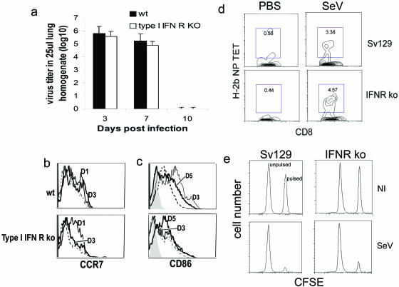
FIG. 2.
Primary immune response against SeV. (a) Virus titers in the lungs of wild-type Sv129 and type I IFN receptor-deficient mice infected intranasally with SeV-52. Error bars indicate standard deviations. (b and c) Analysis of DC maturation (CD86 expression) and migration (CCR7 expression) to the lung draining lymph nodes extracted at different days postinfection; cells gated on the CD11c+ cells. The filled histogram corresponds to the isotype controls. The dashed line corresponds to lymph nodes from mock-infected animals. Continuous lines correspond to lymph nodes extracted from infected animals at the indicated day postinfection (D). Results are representative of two independent experiments. (d) H-2b NP peptide tetramer staining of CD8+ T cells specific for SeV in mice lungs 7 days after infection. (e) In vivo cytotoxicity 5 days postinfection. Lysis of splenocytes pulsed with SeV peptide and labeled with a high dose of CFSE or mock-pulsed and labeled with a low dose of CFSE was evaluated by flow cytometry 18 h after infusion into the infected mice. Results are representative of more than three independent experiments. KO, type I IFN receptor-deficient mice.

