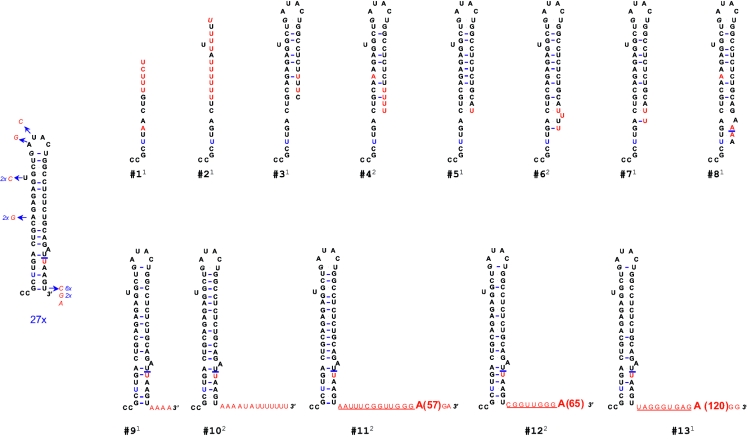
FIG. 2.
Terminal repair of a 5-nt 3′-truncated HCVcon1 replicon. The secondary structure of 3′-SL1 cDNA clones derived from two separate replicon colonies selected from the pFK5.1Δ5 plate (Fig. 1C) is shown. A total of 40 clones were sequenced (20 per colony). Arrows followed by the nucleotide substitution depict mutations found. Shown in red are nucleotides different from the consensus 3′-SL1 (pFK5.1). Underlined sequences indicate the linkers connecting the 3′ end and the poly(A) tail. Clone numbers are indicated under each sequence, and the annotations in superscript (1 or 2) indicate the colony number where it was found.

