FIG.2.
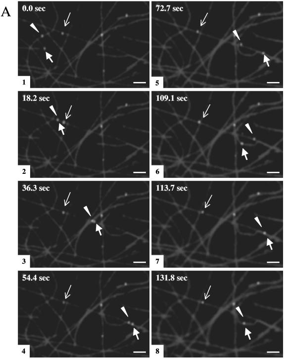
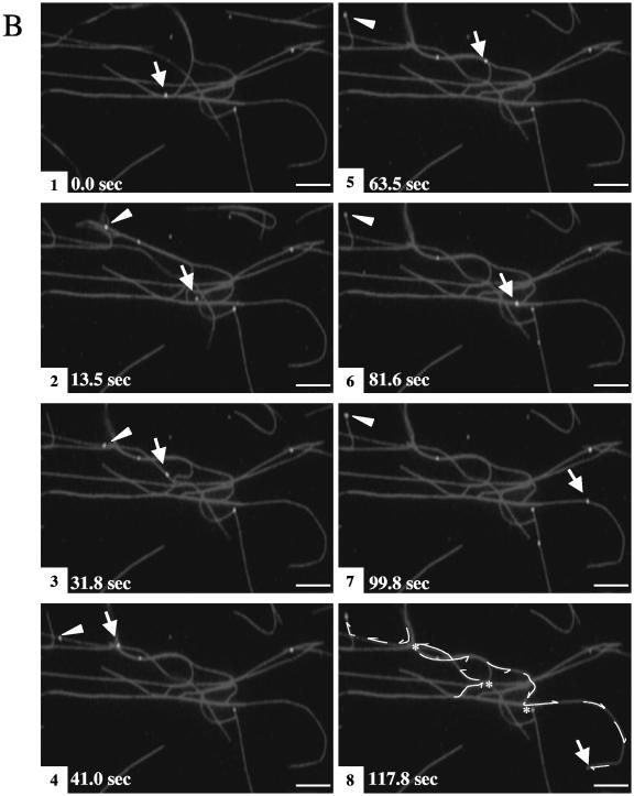
Membrane-associated HSV capsids exhibit ATP-dependent movement along microtubules. Fluorescent HSV-bearing organelles were attached to rhodamine-labeled microtubules as described in the legend of Fig. 1. Following the addition of 500 μM ATP, sequential images were taken using time lapse video microscopy. Eight video frames are shown at selected intervals, as indicated in the upper left corner of each panel. (A) Two particles (thick arrow and arrowhead) move along the microtubules throughout the course of imaging, frequently moving from one microtubule to another at points of intersection, and they occasionally merge (frames 2 and 7) and split apart (frame 3). The average velocity of these particles is ∼0.6 μm/s. A third particle (thin arrow) moves at a much slower rate (an average of ∼0.02 μm/s). (B) One particle (arrow) translocates along microtubules, traveling a total distance of ∼131 μm, switching between microtubules at microtubule junctions (indicated by * in frame 8) and ending its movement at the tip of the microtubule (frame 8). The other particle (arrowhead) stalls after moving ∼22 μm (frame 5). Tracings of these HSV particle movements are shown in frame 8. Bar, 10 μm.


