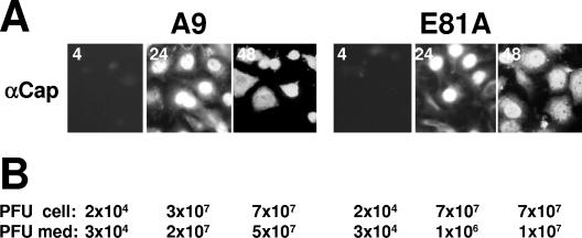
FIG. 8.
Release of progeny virus particles from A9 cells and derivatives expressing dominant-negative CKIIα mutants. (A) Cells grown on spot slides were fixed with paraformaldehyde at 4 h, 24 h, and 48 h p.i. and analyzed by immunofluorescence using a mixture of two mouse monoclonal anticapsid (αCap) antibodies (EIIF3 and B7) and Cy2-conjugated anti-mouse IgGs. (B) Cell-associated infectious virions (PFU cell) and progeny particles released into the medium (PFU med) were determined by standard plaque assays.

