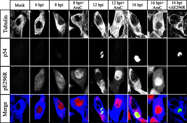
FIG. 6.
Subcellular localization of pE296R protein in ASFV-infected Vero cells by immunofluorescence microscopy analysis. Mock-infected (mock) or ASFV-infected Vero cells were fixed at the indicated times postinfection (hpi) and labeled with anti-tubulin, anti-p54, and anti-pE296R. Antigens were visualized with secondary antibodies coupled to Alexa 697 (tubulin, blue channel), 594 (p54, green channel), and 488 (pE296R, red channel). Cells infected in the presence of AraC were examined at 8, 12, and 16 hpi. A cell infected with the ASFV deletion mutant vΔE296R is also shown. Samples were examined with a Bio-Rad Radiance 2000 confocal laser scanning microscope.

