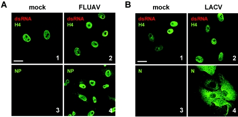
FIG. 4.
Negative-strand RNA viruses. Vero cells were infected at a multiplicity of infection of 5 with FLUAV (A, panels 2 and 4) or LACV (B, panels 2 and 4) or left uninfected (A and B, panels 1 and 3). At 5 h postinfection, cells were fixed and analyzed by immunofluorescence either for dsRNA as indicated in the legend to Fig. 1 (A and B, panels 1 and 2) or for viral antigens (A and B, panels 3 and 4). Bar, 20 μm. All pictures were taken with the same magnification.

