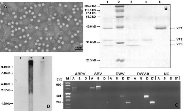
FIG. 1.
Molecular characterization. (A) Electron microscopy image showing empty and filled DWV particles purified from an extract from deformed adult bees. (B) SDS-PAGE of purified DWV. Lane 1, purified virus; lane 2, molecular weight marker; lanes 3 to 5, gel-purified structural proteins. (C) Verification of the purity of DWV preparations by RT-PCR amplification of purified SBV, ABPV, DWV, DWV-it, and negative controls (NC) with primers specific for ABPV (A), SBV (S), and DWV (D and D′). (D) Northern blot of DWV RNA. Total RNA extracted from healthy bees (lane 1) was compared to mRNA (lane 2) and total RNA (lane 3) from infected bees.

