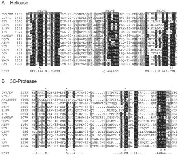
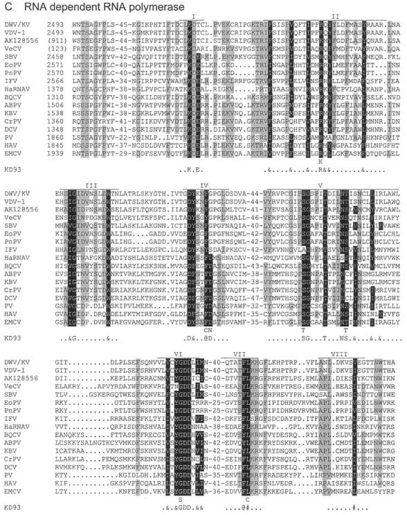
FIG.4.
Protein domain alignments. Shown are alignments of the helicase (A), 3C proteinase (B), and RdRp (C) domains of DWV/KV with those of the other iflaviruses (VDV-1, SBV, VeCV, EoPV, PnPV, and IFV), a human tracheal cDNA clone (AK128556), an ABPV marnavirus (HaRNAV), several (honeybee) cripaviruses (BQCV, KBV, CrPV, and DCV), and three picornaviruses (PV, ECMV, and HAV). The genome location of the first amino acid of each alignment is shown, with those of partial sequences shown in brackets. The lengths of the domains are indicated by the lines above each segment; the conserved amino acids, as identified by Koonin and Dolja (43), are indicated in reverse type and in the line KD93, where “@” refers to an aromatic residue (F, Y, or W); “#” refers to a bulky aliphatic residue (M, I, L, or V); and “&” refers to any bulky hydrophobic residue, aliphatic or aromatic. Other residues that are also conserved among these sequences are shaded in gray.


