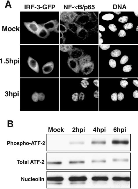
FIG. 2.
Activation of NF-κB, IRF-3, and ATF-2 in RV14-infected cells. (A) Nuclear accumulation of IRF-3 and NF-κB. HeLa cells were transiently transfected with an IRF-3-GFP expression vector and were either mock infected or infected with RV14 and analyzed at 1.5 or 3 hpi. The panels labeled IRF-3-GFP show the localization of the IRF-3-GFP fusion protein, using a fluorescein isothiocyanate filter. The NF-κB/p65 panels show the same field stained with antibodies to detect the p65subunit of NF-κB, visualized using a tetramethyl rhodamine isothiocyanate filter. The DNA panels show the same field stained with Hoechst to reveal cell nuclei. (B) Phosphorylation of ATF-2. Whole-cell lysates (25 μg) prepared from mock-infected cells or cells that had been infected for the indicated amounts of time were analyzed by immunoblotting. ATF-2 was detected by sequentially probing the membrane, using antibodies that recognize the phosphorylated form of ATF-2 (phospho-ATF-2) and all forms of ATF-2 (Total ATF-2). The membrane was also probed with an antibody to nucleolin to show equivalent loading of protein lysates.

