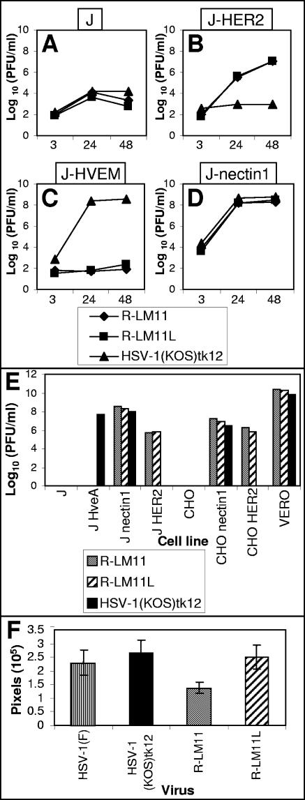
FIG. 6.
Growth and plaque formation of R-LM11 and R-LM11L recombinants. (A to D) Growth curves of R-LM11 and R-LM11L. Replicate cultures of J (A), J-HER2 (B), J-HVEM (C), or J-nectin1 (D) cells were infected with R-LM11 (♦), R-LM11L (▪), or HSV-1(KOS)tk12 (▴) at 10 PFU/cell. Progeny virus was harvested at 3, 24, or 48 h after infection and titrated on Vero cells. (E) Plaque formation of R-LM11 and R-LM11L. R-LM11 (gray bars), R-LM11L (hatched bars), and HSV-1(KOS)tk12 (black bars) were plated in the indicated cell lines. Monolayers were fixed at 24 or 48 h after infection, and plaques were visualized by X-Gal or Giemsa staining. (F) Plaques formed as shown in panel E were photographed, and the plaque areas were measured by means of the Histogram program and expressed as pixels. For each virus, the areas of at least 20 plaques were measured. Histograms represent averages; error bars, standard deviations.

