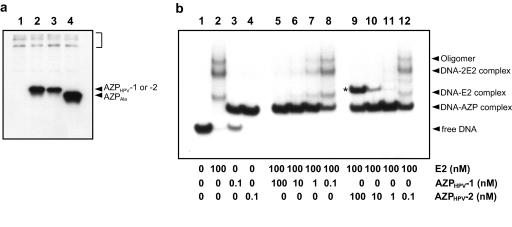
FIG. 5.
Identification of the cause of more efficient inhibition of HPV-18 DNA replication by AZPHPV-2. (a) Western blot analysis of AZPs. The immunoblot was obtained by using protein extracts derived from the transient replication assays performed for Fig. 4c. The AZP expression plasmids used were pCMV-AZPHPV-1 (lane 2), pCMV-AZPHPV-2 (lane 3), and pCMV-AZPAla (lane 4). When pcDNA3.1 (lane 1) was used as the control, there was no signal except for faint artifact signals derived from endogenous proteins cross-reactive to the anti-T7-tag antibody (shown by the square bracket). (b) Competitive binding experiment with E2 and AZP. DNA-binding assays were performed with the 32P-labeled 56-bp probe containing E2BS-3 and E2BS-4 along with a constant level of E2 protein (100 nM) in the presence of increasing concentrations of AZP (nM), from lane 8 to lane 5 for AZPHPV-1 and from lane 12 to lane 9 for AZPHPV-2. Lanes 1 to 4 indicate band positions of free DNA, DNA bound to E2, DNA bound to AZPHPV-1, and DNA bound to AZPHPV-2, respectively, which are used as markers for lanes 5 to 12. The concentrations of AZP and E2 protein used are indicated below each lane. The identities of the shifted bands are described in Results and Discussion.

