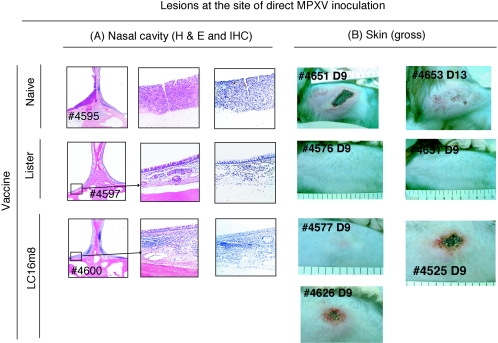
FIG. 4.
Histology of the nasal cavity caused by intranasal challenge with MPXV strain Liberia (A) and macroscopic lesions at the site of subcutaneous inoculation with MPXV strain Zr-599 (B). The identification numbers of monkeys are given. Analyses by H&E staining (A, low and high magnifications) revealed that the lesions of naïve monkey 4595 were characterized by destruction of mucous membrane structures, disappearance of mucosal epithelial cells resulting in ulcer formation, necrosis, and hyperplasia. MPXV antigens were present in the lesions. In contrast, the mucous membranes of the nasal cavity into which MPXV was inoculated in Lister-immunized monkey 4597 were normal, and MPXV antigens were not detected. Although the mucous membranes of the LC16m8-immunized monkey 4600 showed infiltration of inflammatory cells, the structure was maintained without necrosis. Furthermore, MPXV antigens were not detected. (B) Erythematous, vesicular, and ulcerative lesions appeared in the SC-Naïve group. The maximum diameter of the lesions exceeded 10 cm on day 14 postchallenge. In the SC-LC16m8 group, similar but milder lesions were observed, while no obvious lesions were detected at the site of inoculation in the SC-Lister group.

