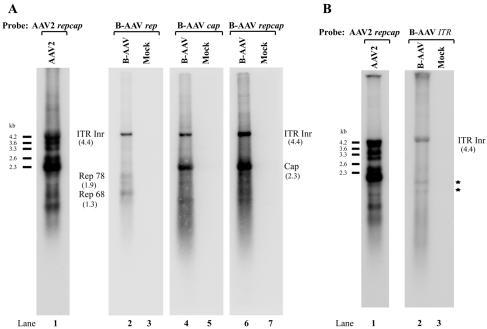
FIG. 3.
Northern blot analysis of B-AAV RNA. Total RNA was isolated 36 to 40 h postinfection of MDBK cells or from uninfected MDBK cells (Mock), and mRNA purified from 10 μg of total RNA was used for Northern analysis. The blot was hybridized to either a whole B-AAV repcap probe (A, lanes 2 and 3), rep probe (A, lanes 4 and 5), cap probe (A, lanes 6 and 7), or TR probe (B, lanes 2 and 3), which are diagrammed in Fig. 2B and described in Materials and Methods. The identities of bands protected by B-AAV RNA are shown with their respective sizes in parentheses. AAV2 RNA from AAV2-infected 293 cells was used as size markers (A and B, lane 1). Bands indicated with an asterisk (B, lane 2) were likely degraded RNAs.

