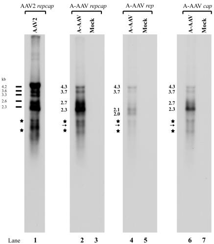
FIG. 6.
Northern blot analysis of A-AAV RNA. Total RNA was isolated 36 to 40 h postinfection from A-AAV-infected chicken primary kidney cells. mRNA purified from 10 μg of total RNA was subjected to Northern analysis; uninfected mRNA was used as a mock control. The Northern blot was hybridized with either a whole A-AAV repcap probe (lanes 2 and 3), a rep probe (lanes 4 and 5), or a cap probe (lanes 6 and 7), which are diagrammed in Fig. 5. The sizes of different A-AAV RNA species are shown. AAV2 RNA from AAV2-infected 293 cells was used as size markers. Because the bands marked with an asterisk appear with all probes, it is likely that they are degradation products. P19-generated RNAs polyadenylated at (pA)p would be expected to run in this region, and a putative band detected with the repcap and rep probe, but not with the cap probe, is marked with an arrow; however, as described in the text, the appearance of degradation products in this area of the gel precluded their accurate assessment.

