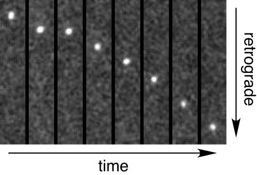
FIG. 2.
VP26-null capsid retrograde axonal transport. Example of virus particle transport resulting from infection of dorsal root sensory neurons with PRV-GS1205 and imaged within the first hour postinfection. A montage of eight frames from a subregion of a time-lapse recording are shown (see movie M1 in the supplemental material for the entire time-lapse recording). Each frame is a 200-ms exposure representing every fourth frame of the original recording (the montage represents a 6.4-s time window). A single VP26-null capsid (mRFP1-VP1/2) complex is shown in the montage. The frames are each 2.7 μm × 15.2 μm.

