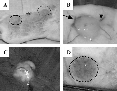
FIG. 3.
Gross appearance of collagen scaffolds containing M. bovis BCG in BALB/c mice. (A) Gross macroscopic appearance of mouse with subcutaneous implants 1 week after implantation (circled areas). (B) The skin flap reveals the implant adhering to the underside of the skin, and arrows denote vessels surrounding the pellet that were typically observed as early as 1 week postimplantation. (C) Implants in situ for 3 months revealed possible caseous necrosis. (D) Vascular footprint (circle) that remains after removal of gel implant.

