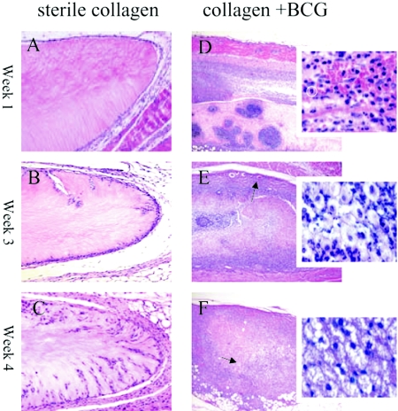
FIG. 4.
Histological sectioning (H&E staining) of collagen scaffolds implanted subcutaneously in mice for 1, 3, and 4 weeks. Panels A to C (magnification, ×100) represent implanted collagen scaffolds that did not contain M. bovis BCG that were in situ for 1 week (A), 3 weeks (B), and 4 weeks (C). Little cellular recruitment into the collagen scaffolds was observed compared to that shown in panels D to F (magnification, ×50) with their respective insets (magnification, ×630 using water immersion objective), which represent BCG-containing scaffolds that were implanted for 1, 3, and 4 weeks, respectively. Massive cellular recruitment is marked by the abundance of hematoxylin-stained nuclei throughout the gel (D to F). After 1 week (D), mononuclear cells predominated (typical neutrophil circled), whereas at 3 weeks (E) and 4 weeks (F), macrophages became the dominant cell population and possible lymphocytes began to appear. Vascularization of the fibrous capsule was evident from week 3 (arrow in panel E). After 4 weeks, an eosinophilic acellular pattern was observed in the infected scaffolds (arrow in panel F). A lower magnification was used for BCG-containing scaffolds to capture the full extent of inflammation, and a higher magnification was used to reveal individual cell types of the infiltrate.

