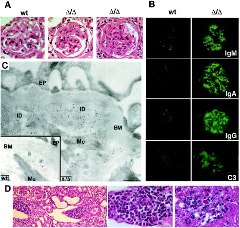
Figure 3.
Immune complex glomerulonephritis in the absence of α-mannosidase II. (A) In comparison with glomeruli from wild-type mice (Left), thickening of the mesangium is observed with capillary lumen obstruction in mutant mice (Center and Right). (Hematoxylin/eosin stain of glomeruli at ×100 magnification is shown.) (B) The glomeruli of mice lacking α-mannosidase II contain high levels of Ig deposits composed of IgM, IgA, and IgG [the latter include IgG1, IgG2a, and IgG2b (not shown)], as well as complement component C3 (×400 magnification shown). Antibody deposits were also noted in other tissues, including lung and liver, but to a lesser extent (not shown). (C) Immunogold labeling of a glomerulus from a mutant mouse, showing immune deposits (ID) between the glomerular basement membrane (BM) and mesangial cell processes (Me). Gold particles indicate the presence of IgG/IgM in the ID. (Inset) Similar field from a wild-type mouse, showing few gold particles. EP, Foot processes of glomerular epithelium (bar, 0.5 μm). (D) Mononuclear leukocytic infiltrates in kidney (Left and Center) and liver of mutant mice (×400): lymphocytes, plasma cells, and neutrophils. Infiltrates in liver (Right) are found with evidence of hepatocyte degeneration and accumulated bile. Results shown are from mice that are either 8 months (A, B, and D) or 13 months (C) of age.

