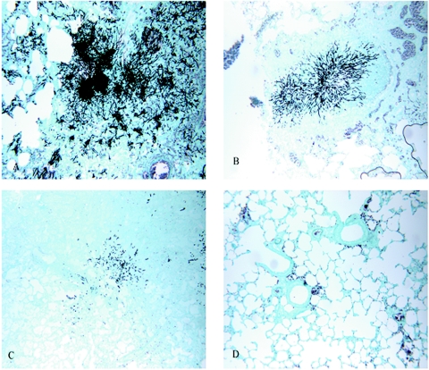
FIG. 2.
Sections of lungs from immunosuppressed rats infected with invasive A. fumigatus (Grocott-Gomori methenamine-silver nitrate stain; magnification, ×100). (A) Untreated lung tissue from a control infected rat showing severe granulomatous pneumonia and large numbers of branching fungal hyphae; (B) lung tissue from an infected rat treated for 7 days with EDTA alone showing mild consolidation of the parenchyma and accumulation of fungal hyphae; (C) lung tissue from an infected rat treated for 7 days with ABLC alone showing mild pneumonia and an abundance of organisms; (D) lung tissue from an infected rat treated for 7 days with ABLC plus EDTA showing no significant lesions and no organisms.

