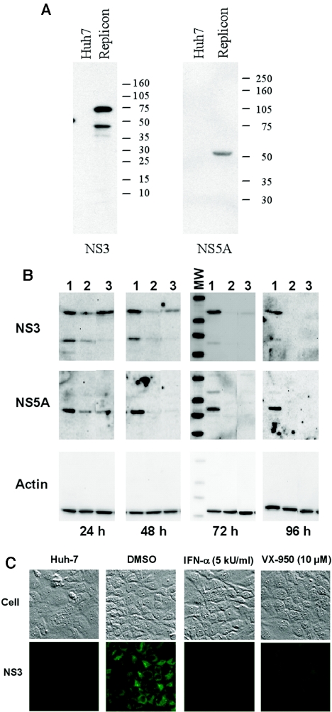
FIG. 3.
Reduction of HCV proteins in the replicon cells by VX-950. (A) Specificity of the antibodies against HCV NS3 and NS5A proteins in Western blot analysis. Cell lysates from the parental Huh-7 (Huh7) or HCV replicon (Replicon) cells were analyzed by SDS-PAGE, transferred, and probed with specific antibody against NS3 (left) or NS5A (right) antigens. The molecular weight markers are indicated at the right. (B) Reduction of HCV proteins in a Western blot. Replicon cells were incubated with 0.2% DMSO (no-compound control) (lane 1), 5 kU/ml IFN-α (lane 2), or 10 μM VX-950 (lane 3) for 24, 48, 72, or 96 h. Equal amounts of protein from each cell extract were subjected to SDS-PAGE and subsequent Western blot analysis with specific antibodies against NS3 (top), NS5A (middle), or β-actin (bottom). The molecular weight markers (MW), shown in the left lane of the 72 h subpanel, are the same as those used in panel A. (C) Reduction of HCV proteins in immunofluorescence staining. The parental Huh-7 cells (left) or HCV replicon cells were either incubated with 0.2% DMSO (no-compound control), 5 kU/ml IFN-α, or 10 μM VX-950 for 72 h and then subjected to regular microscopy (top) or to immunofluorescence staining using an anti-NS3 antibody (bottom).

