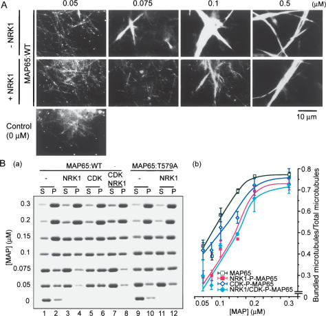
Figure 5.
Suppression of the MT-bundling activity in vitro of NtMAP65-1a upon phosphorylation by NRK1/NTF6 MAPK. (A) Dark-field micrographs of bundled MTs after bundling had been promoted by His-T7NtMAP65-1a (MAP65:WT). Taxol-stabilized MTs were incubated with MAP65:WT (top) or NRK1-phosphorylated MAP65:WT (middle) at the indicated concentrations. The bottom panel shows the distribution of MTs in the absence of NtMAP65-1a. (B) Quantification of bundled MTs promoted by NtMAP65-1a. (Panel a) Staining by Coomassie Brilliant Blue of tubulin in bundled and unbundled MTs after SDS-PAGE. Bundled MTs with His-T7-NtMAP65-1a and individual MTs were separated by low-speed centrifugation. Aliquots of supernatants (S), which included nonbundled MTs and pellets (P) that included bundled MTs, were subjected to SDS-PAGE and then the gel was stained. (Panel b) Graphs showing ratios of bundled MTs to total MTs at various concentrations of His-T7-NtMAP65-1a (MAP65:WT). Relative amounts of MTs that had been bundled by MAP65:WT (open squares), NRK1-phosphorylated proteins (red filled squares), CDK-phosphorylated proteins (open diamonds), and NRK1 plus CDK-phosphorylated proteins (blue filled diamonds) were calculated from the results of cosedimentation assays after low-speed centrifugation. Independent assays were performed more than three times, and average values with standard deviations are shown.

