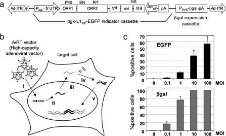
Fig. 1.
Outline of the adenovirus/retrotransposon hybrid vector (A/RT vector) system. (a) Schematic structure of the A/RT vector. The A/RT vector is a high-capacity, helper-dependent adenovirus vector. The sequences composing the A/RT vector are, from left to right: the 5′ inverted terminal repeat (Ad-ITR) of human adenovirus type 5 including the packaging signal (ψ), the L1RP-EGFP cassette, a β-gal marker expression cassette, and the 3′ Ad-ITR. The pgk-L1RP-EGFP cassette consists of a mouse phosphoglycerate kinase promoter-1 (Ppgk), the 5′ UTR, and two ORFs [ORF1, P40; ORF2, endonuclease (EN)/RT] of L1RP, the EGFP transgene, and a polyadenylation signal (pA). The EGFP transgene cloned into the 3′ UTR of L1RP in the reverse orientation (PFGE) is flanked by the CMV promoter (PCMV) and inverted pA sequences also in the reverse orientation and is interrupted by a forward-splicing intron (IVS) (13, 14). The structure of the splice-interrupted EGFP indicator gene cassette before and after L1 retrotransposition is shown in Fig. 2c. This A/RT vector construct can be introduced into cells via direct transfection in the form of plasmid DNA or via infection after being packaged into an adenovirus. (b) Two-stage transduction with the A/RT virus. The A/RT virus infects target cells efficiently as an adenovirus (i). A full-length active L1 element is transcribed from the constitutively active pgk promoter to produce bicistronic mRNA (ii). The RNA undergoes processing and is exported from the nucleus (iii). In the cytoplasm, the ORF1 and ORF2 proteins are translated and specifically function on the RNA that transcribed them (cis preference) (6, 7). The L1 RNA molecule, ORF1, and ORF2 proteins assemble into a ribonucleoprotein complex that is an intermediate in retrotransposition (iv) and then imported back into the nucleus, where the L1 RNA is reverse transcribed and integrated into the host genome for stable gene expression (v) (2). (c) Retrotransposition after A/RT virus infection. Gli36 cells were infected with the A/RT virus at different mois. Seven days after infection, the cells were analyzed for expression of β-gal from the adenovirus backbone and EGFP from retrotransposition events. Data shown are averages and SDs from experiments performed in quadruplicate.

