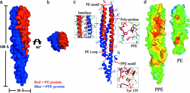
Fig. 3.
Crystal structure of the M.tb. PE/PPE protein complex. (a) Surface representation of the PE/PPE protein complex. The PE protein Rv2431c is shown in red, and the PPE protein Rv2430c is in blue. (b) The PE/PPE protein complex viewed down its longitudinal axis. (c) Ribbon diagram of the PE/PPE protein complex. The complex is composed of seven α-helices. Two α-helices of the PE protein interact with two helices of the PPE protein to form a four-helix bundle. Regions of high sequence conservation are indicated by arrows and discussed in the text. (d) Interface hydrophobicity of the PPE and PE proteins. The hydrophobicity of the interaction interface between the PPE and PE protein is color-coded: the most apolar regions are indicated in red, orange, and yellow, and the most polar regions are indicated in blue. Notice the extensive apolar regions that are shielded from solvent as the complex forms.

