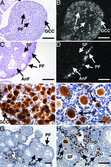
Fig. 1.
Sohlh1 mRNA and protein expression. (A–D) In situ hybridization with Sohlh1 riboprobe on newborn (A and B) and 6-week-old (C and D) ovarian tissue. Bright-field (A and C) and dark-field (B and D) views are shown. Arrows throughout show locations of germ cell cysts (GCC), primordial follicles (PF), primary follicles (PrF), secondary follicles (SF), and antral follicles (AnF). Sohlh1 transcripts localize mainly to germ cell cysts and oocytes in primordial follicles. (E–H) Rabbit antibodies against SOHLH1 were used to perform immunohistochemistry on newborn (E), 9-day-old (F), and 6-week-old (G and H) ovaries. Immunoreactivity to the SOHLH1 protein stained brown, and cell nuclei were counterstained blue with hematoxylin. (Scale bars: A–D and G and H, 100 μm; E and F, 20 μm.)

