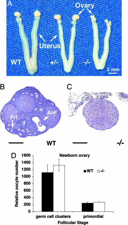
Fig. 2.
Sohlh1 adult knockout anatomy, histology, and histomorphometric analysis. (A) Gross reproductive tracts dissected from WT, heterozygous (+/−), and homozygous (−/−) Sohlh1 mice. Note markedly smaller ovaries in Sohlh1−/− mice. (B) WT ovary with advanced antral follicle (AnF) as well as primary follicles (Prf). (C) Sohlh−/− ovary (−/−) lacks germ cells. (D) Five pairs of newborn ovaries from WT and Sohlh1 knockout (−/−) mice were sectioned, and oocytes within the germ cell clusters and primordial follicles were counted. No significant differences were observed between the WT and knockout ovaries. Data are represented as mean values, with error bars representing the SEM. Fisher's exact t test was used to calculate P values. (Scale bars: 400 μm.)

