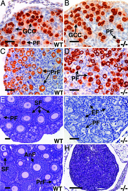
Fig. 3.
Sohlh1 knockout histology and immunohistochemistry; WT and knockout (−/−) data are shown. (A and B) Newborn ovaries stained with antibodies against GCNA1 show no difference in primordial follicles (PF) or germ cell cysts (GCC) between WT (A) and knockout (B). (C and D) Three-day ovaries stained anti-MSY2. Primary follicles (PrF) are seen in WT (C) but not knockout (D) ovaries. (E and F) Periodic acid/Schiff reagent (PAS) staining of 7-day WT (E) and knockout (F) ovaries show fewer follicle types and empty follicles (EF) in the knockout (F). (G and H) PAS staining of 3-week ovaries shows no remaining oocytes in the knockout (H) but all stages of development in the WT (G). (Scale bars: A–G, 50 μm; H, 400 μm.)

