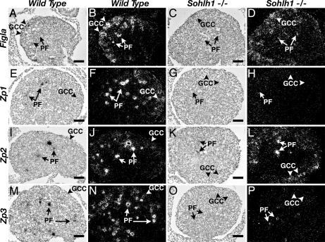
Fig. 4.
Expression of Figla, Zp1, Zp2, and Zp3 in WT and Sohlh1−/− ovaries. Bright-field (A, C, E, G, I, K, M, and O) and their corresponding dark-field (B, D, F, H, J, L, N, and P) images of in situ hybridization from WT (A, B, E, F, I, J, M, and N) and Sohlh1−/− (C, D, G, H, K, L, O, and P) newborn ovaries. In newborn WT ovaries, Figla (A and B), Zp1 (E and F), Zp2 (I and J), and Zp3 (M and N) are expressed in germ cell cysts (GCC; arrowheads) and primordial follicles (PF; arrows). Expression of Figla (C and D) and Zp2 (K and L) are detectable in Sohlh1−/− ovaries by in situ hybridization. Zp1 (G and H) and Zp3 (O and P) are not detectable by in situ hybridization in Sohlh1−/− ovaries. Magnification is the same in A–P. (Scale bars: 40 μm.)

