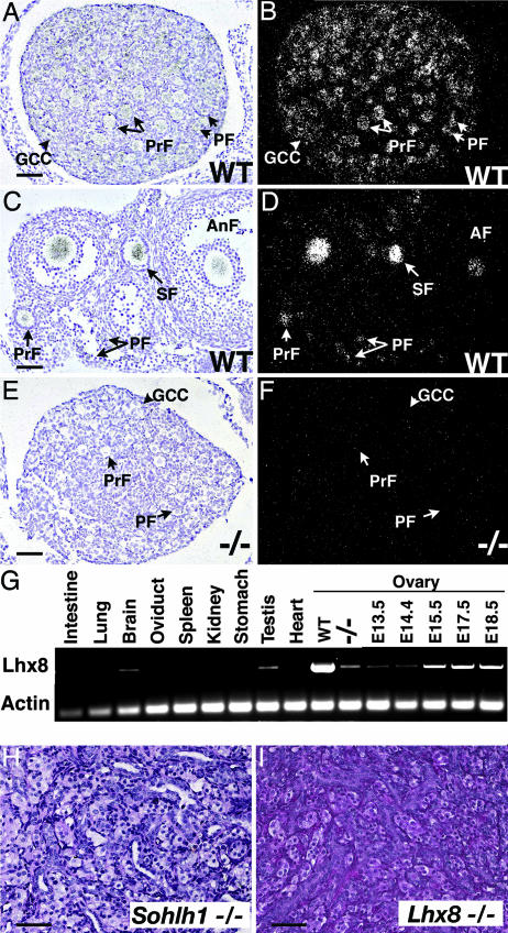
Fig. 5.
Lhx8 expression and ovarian phenotype. (A–F) Bright-field (A, C, and E) and dark-field (B, D, and F) views of in situ hybridization are shown with Lhx8 riboprobe to WT newborn (A and B) and 6-week-old (C and D) ovaries. (E and F) The Lhx8 riboprobe showed no significant hybridization to Sohlh1−/− ovaries. (G) Oligonucleotides corresponding to Lhx8 amplified RNA in WT testes and newborn ovaries (WT) but showed a dramatic decrease in Sohlh1−/− ovaries (−/−). Total RNA from embryonic ovaries (E13.5, E14.5, E15.5, E17.5, and E18.5) was isolated and also amplified with Lhx8-specific primers. (H and I) Periodic acid/Schiff reagent staining of 12-week old (adult) ovaries from Sohlh1−/− (H) and Lhx8−/− (I) shows a lack of germ cells in both mutants. GCC, germ cell cyst; PF, primordial follicle; PrF, primary follicle; SF, secondary follicle; AnF, antral follicle. (Scale bars: 40 μm.)

