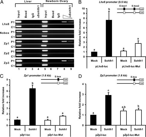
Fig. 6.
SOHLH1 binding and transactivation of the Lhx8, Zp1, and Zp3 promoters. (A) ChIP assays with anti-SOHLH1 antibodies on newborn ovary and liver extracts. Anti-SOHLH1 antibodies precipitate genomic DNA containing conserved E boxes from Lhx8, Zp1, and Zp3 promoter regions (P1) but not control genomic regions (C1). PCR amplifications of the E boxes are described in the legend of Fig. 12 and Table 2. “Input” is PCR product from chromatin pellets before immunoprecipitation. Samples incubated with anti-SOHLH1 antibody (αSohlh1) and the control sample without antibody (Bead) or IgG were used as templates for PCR. (B–D) Transient transfection analyses of Lhx8 (B), Zp1 (C), and Zp3 (D) promoter regions with SOHLH1. Reporter constructs containing the WT (filled box, pLhx8-luc, pZp1-luc, and pZp3-luc) or mutant E boxes (open box, pLhx8-luc-Mut, pZp1-luc-Mut, and pZp3-luc-Mut) were cotransfected with vector expressing SOHLH1 or the empty vector (Mock). Lhx8 putative promoter contains two E boxes, and both were mutated. Conserved E boxes and mutated sequences are shown in Fig. 12 and Table 1. The mean fold increase in luciferase activity (± SEM) of triplicate experiments relative to the empty vector is shown. Statistical significance was determined by one-way ANOVA followed by the Tukey–Kramer honestly significant difference test for multiple comparisons. Bars marked with difference letters (a, b, and c) indicate statistical significance (P < 0.001).

