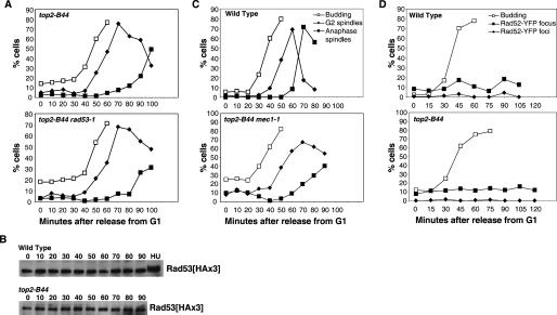
Figure 2.
G2/M delay in top2-B44 is DNA damage checkpoint-independent. (A,C) Cell cycle analysis of double mutants of top2∷KAN pCEN-top2-B44 combined with DNA damage checkpoint mutants, performed as described in Figure 1 after G1 synchrony. (A) Cell cycle progression after release from G1 in top2-B44 rad53-1. (C) Cell cycle progression after release from G1 in top2-B44 mec1-1 sml1Δ. (B) Western blot showing Rad53 phosphorylation shift after hydroxyurea (HU) treatment, but no shift in wild-type or top2-B44 cells progressing through the cell cycle at 32°C, released from mating pheromone-induced G1 synchrony. (D) Rad52 foci (a measure of the presence of DNA breaks) in wild-type or top2-B44 cells progressing through the cell cycle at 32°C, released from mating pheromone-induced G1 synchrony. Foci = cells with more than one fluorescent dot; focus = cells with one fluorescent dot.

