Fig. 4.
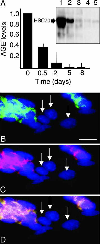
AGE modification in undifferentiated and differentiated ES cells. (A) AGE levels in undifferentiated ES cells (day 0) and cells that have been triggered to differentiate by the removal of LIF. Inset shows a Western blot demonstrating an almost exclusive AGE modification of one single protein, which was identified as HSC70. AGE modification of HSC70 is shown after 0 (lane 1), 0.5 (lane 2), 2 (lane 3), 5 (lane 4), and 8 (lane 5) days of differentiation. Error bars represent standard error of at least three measurements. (B) Overlay of AGE immunodetection (green) and DAPI (blue) signals. (C) Overlay of carbonyl (red) and DAPI signals. (D) Overlay of AGE, carbonyl, and DAPI signals. In B–D, arrows indicate cells that are negative for both carbonyl and AGE staining. Representative images are shown. (Scale bar, 25 μm.)

