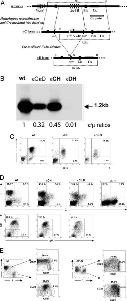
Fig. 1.
Ig transgenic κLC expression by splenocytes from mutant animals. (A) Structure of the targeted locus. At the top is shown the WT κ locus and the 3′ probe. In the middle is shown the structure of the κC mutated locus after targeted recombination and neoR deletion. A pVH promoter and the human VκJκ exon replaced the Jκ cluster. Dashed lines mark homologous sequences used as 5′ and 3′ arms of the construct. At the bottom is shown the κD mutation after cre-deletion of all knockin elements. B, BamHI; Bs, BsmI; S, SacI. (B) Northern blot and QPCR analysis of κ mRNAs. RNA (10 μg) from spleen of WT, κCH, κC/κD, and κDH mice were analyzed on 1% agarose/0.7 M formaldehyde gels and hybridized with a Cκ probe. The 1.2-kb band corresponding to normal-size κ mRNA is indicated (arrow). κ/μ ratios were calculated by QPCR. (C) Flow cytometric analysis of κLC-expressing cells in spleen. WT, κCH, or κCκD splenocytes were stained as indicated. Numbers indicate the percentages of cells in the corresponding quadrants (from one representative of five independent experiments). (D) Flow cytometric analysis of WT, κCH, κCκD, or κDH splenocytes stained as indicated. Numbers indicate the percentages of total cells in the corresponding quadrants (from one representative of five independent experiments). (E) CD43-enriched B cells from WT and κCκD mice were analyzed in two gates corresponding to κ- and λ-only B cells for WT mice or κ-only and κ/λ B cells for κCκD mice; these two gates were then analyzed for CD23 and CD21 expression.

