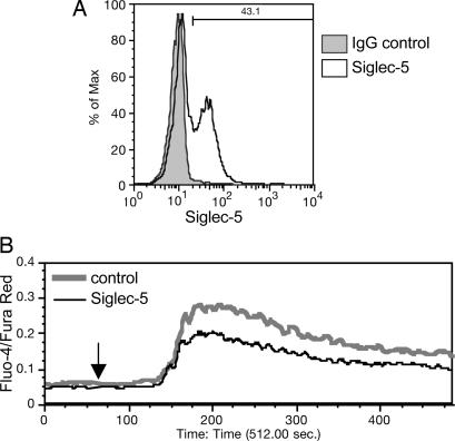
Fig. 7.
Siglec-5 expression on Jurkat T cells inhibits anti-CD3-induced intracellular calcium mobilization. Jurkat cells were mock-transfected with no DNA or transfected with 2 μg of pSig5 per 2 × 10⁶ cells using the Nucleofector device. After 24 h, Siglec-5 expression was analyzed by flow cytometry with anti-Siglec-5 and goat anti-mouse IgG Alexa Fluor 488, by using a nonspecific mouse IgG as background (A). The percentage of Siglec-5-positive cells is indicated. Transfected cells were loaded with calcium-sensing dyes, Fluo-4 and Fura Red, and then analyzed for responses to soluble anti-CD3 by real-time flow cytometric analysis (B). The arrow at 60 s indicates the time of mAb addition. The experiment was repeated twice with different transfectant Jurkat cells and produced similar results.

