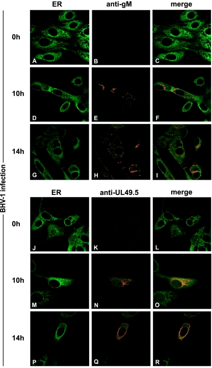
FIG. 7.
Subcellular redistribution of UL49.5 and gM in BHV-1-infected MDBK cells, analyzed by confocal laser scanning microscopy. MDBK cells were infected with BHV-1 at an MOI of 1. gM was detected in infected cells with gM-specific rabbit antibodies at the indicated times postinfection (B, C, E, F, H, and I). UL49.5 was detected with UL49.5-specific rabbit antibodies (K, L, N, O, Q, and R). The ER/cis-Golgi was stained with concanavalin A-Alexa 488 (A, C, D, F, G, I, J, L, M, O, P, and R). The anti-gM and anti-UL49.5 rabbit antibodies were visualized with Alexa 594-conjugated goat anti-rabbit IgG. In the overlays, colocalization is shown in yellow.

