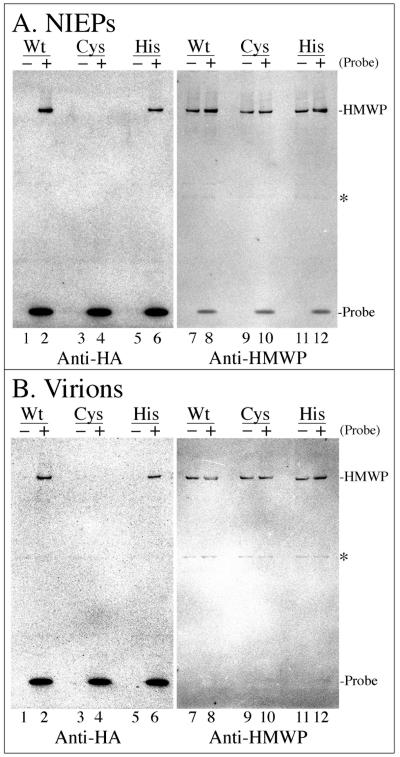
FIG. 6.
DUB probe binds to wild-type and H162A mutant HMWP, but not to C24I mutant HMWP, in virions and NIEPs. Samples of the virions and NIEPs shown in Fig. 5 were reacted with (+) or without (−) the DUB probe, subjected to SDS-PAGE (4 to 12% gel with MOPS electrode buffer), and analyzed by phosphorimaging following Western immunoassay with antibodies to detect the DUB probe (anti-HA) or to detect HMWP (anti-UL48). Shown here are images of one membrane with proteins from the NIEP preparations (A) and a second membrane with proteins from the virion preparations (B). Each membrane was probed first with anti-HA and imaged, rehydrated for 10 min in TN+BSA, and then probed with anti-UL48 and imaged again. Abbreviations to the right of images are as in Fig. 2; asterisks indicate the position of a ≈63-kDa cross-reacting band that is not probe specific. Amounts of DUB probe bound relative to the amount of HMWP present were calculated using measurements from the four phosphorimages shown. Values for wild-type particles were determined as the quotient of the HMWP intensities in lane 2 divided by that in lanes 7 plus 8; values for H162A particles were determined as the quotient of the HMWP intensities in lane 6 divided by that in lanes 11 plus 12.

