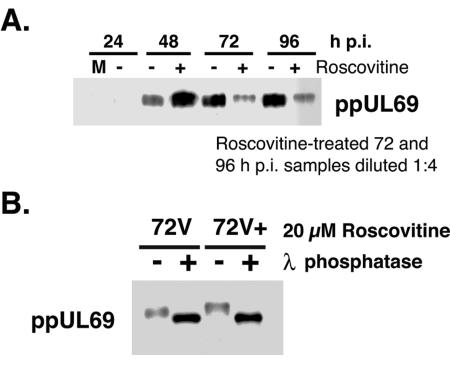
FIG. 6.
ppUL69 accumulates in a hyperphosphorylated form in the presence of Roscovitine. (A) Samples were processed for Western blotting as described in the legend to Fig. 3. To distinguish the phosphorylated forms of ppUL69, lysates were separated by SDS-PAGE in gels containing 4.2% cross-linking. Lanes were loaded by equivalent cell numbers except for drug-treated samples from 72 and 96 h p.i., which were diluted to prevent saturation of the chemiluminescent signal. +, treated with Roscovitine; −, treated with DMSO as a control; M, mock-infected cells. (B) Phosphatase treatment of infected-cell proteins. Cell pellets were extracted as described in Materials and Methods, and lysates were treated with lambda phosphatase for 30 min (+). The reaction was stopped by adding an equivalent volume of 2× RSB followed by boiling of the samples. Control samples (−) were processed in parallel. Proteins were separated by SDS-PAGE as described above. The filter was reacted with antibody specific for ppUL69. 72V, untreated viral samples from 72 h p.i.; 72V+, roscovitine-treated viral samples from 72 h p.i.

