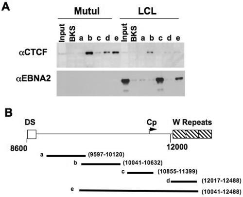
FIG. 1.
DNA affinity pull-down of CTCF and EBNA2 with EBV DNA. (A) Western blots of CTCF and EBNA2 binding to different biotinylated DNA fragments of EBV using nuclear extracts from MutuI (type I EBV-positive cell line) and LCL3456 (type III EBV-positive cell line). Inputs represent 10% of total nuclear proteins used for DNA affinity. BKS is a random biotinylated DNA fragment of pBluescript KS+. Fragments a to e represent DNA affinity using different DNA segments of EBV encompassing a region between the DS and W repeats of EBV. αCTCF and αEBNA2, anti-CTCF and anti-EBNA2 antibodies, respectively. (B) Schematic diagram of the different DNA segments of EBV used in the DNA affinity assay. The segments are labeled a to e, and the regions of EBV covered by each segment are labeled with its EBV coordinates.

