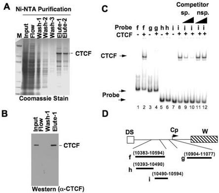
FIG. 2.
Electrophoretic mobility shift assay of CTCF with EBV DNA. (A) Coomassie gel of Ni-NTA purification of His-tagged CTCF expressed in Sf9 insect cells. Sf9 cell extracts expressing His-tagged CTCF were loaded onto Ni-NTA beads, washed with 20 mM imidazole buffer, eluted with 250 mM imidazole elution buffer, and dialyzed in PBS containing 20% glycerol. (B) Western blot of His-tagged CTCF. Different fractions of proteins during the purification process were electrophoresed on an 8 to 16% SDS-PAGE gel, transferred onto nitrocellulose membrane, and probed using a rabbit polyclonal anti-CTCF antibody (α-CTCF). (C) Autoradiogram of in vitro EMSA showing CTCF shifting of EBV DNA probes. Purified His-tagged CTCF was used for gel shift of various EBV 32P-labeled DNA probes. Lanes 1 and 2 represent EMSA with probe f, lanes 3 and 4 are EMSA with probe g, lanes 5 and 6 are EMSA with probe h, and lanes 7 to 12 are EMSA with probe i. Lanes 9 to 12 represent EMSA with specific (sp.) and nonspecific (nsp.) cold DNA competitors at 10- and 100-fold excess. (D) Schematic diagram of various EBV DNA probes used in EMSA. The probes cover regions upstream of the C promoter, between EBV coordinates 10383 to 10594 and 10904 to 11077.

