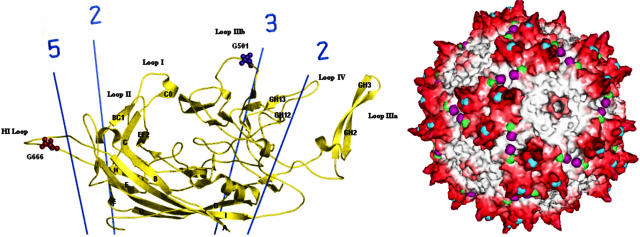
FIG. 1.
(Left) Sequence motifs characteristic of glycosaminoglycan modification are shown as spheres on the part of the capsid subunit structure that is common to VP1, VP2, and VP3 (58). Asn496 to Ser498 and Asn519 to Ser521 are on outer loops near the three- and five-fold axes respectively. (Right) In the assembled capsid, these sequences are in prominent positions on the outer surface. Asn496 to Ser498 are shown in blue near the tips of protrusions that surround each three-fold axis. Asn519 to Ser521 are shown in green near the five-fold axes.

